A new optical ultrasound needle developed by researchers at UCL and Queen Mary University of London (QMUL) is allowing soft tissues inside the patient to be imaged in real-time. The team has been developing this technology in a clinical setting for over four years.
Keyhole surgery?
Minimally invasive surgery, or keyhole surgery, is a surgical technique that operates on the target location using a small incision located elsewhere in the body.
Currently, doctors are relying on external ultrasound probes and pre-operative imaging scans to visualize the soft tissues.
This has limits on the resolution, location, and differentiation of soft tissues.
What does this new ultrasound imaging provide?
This new ultrasound imaging needle provides a high-resolution image of 64 microns (equal to 9 red blood cells), up to 2.5cm in front of the instrument, inside the patient’s body.
This allows a detailed differentiation and real-time view of the soft tissues, without harm to the patient.
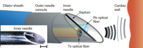
The optical ultrasound needle functions using miniature optical fibers that deliver a brief pulse of light. This light generates ultrasonic pulses that are reflected by tissues. The reflections are detected by a sensor on the second optical fiber, resulting in real-time images.
This new optical ultrasound needle can be used on needle tips under 1mm. It is good for small tissue targets and has potential application in guiding epidural needles.